Uterine Fibroids: Symptoms & Diagnosis
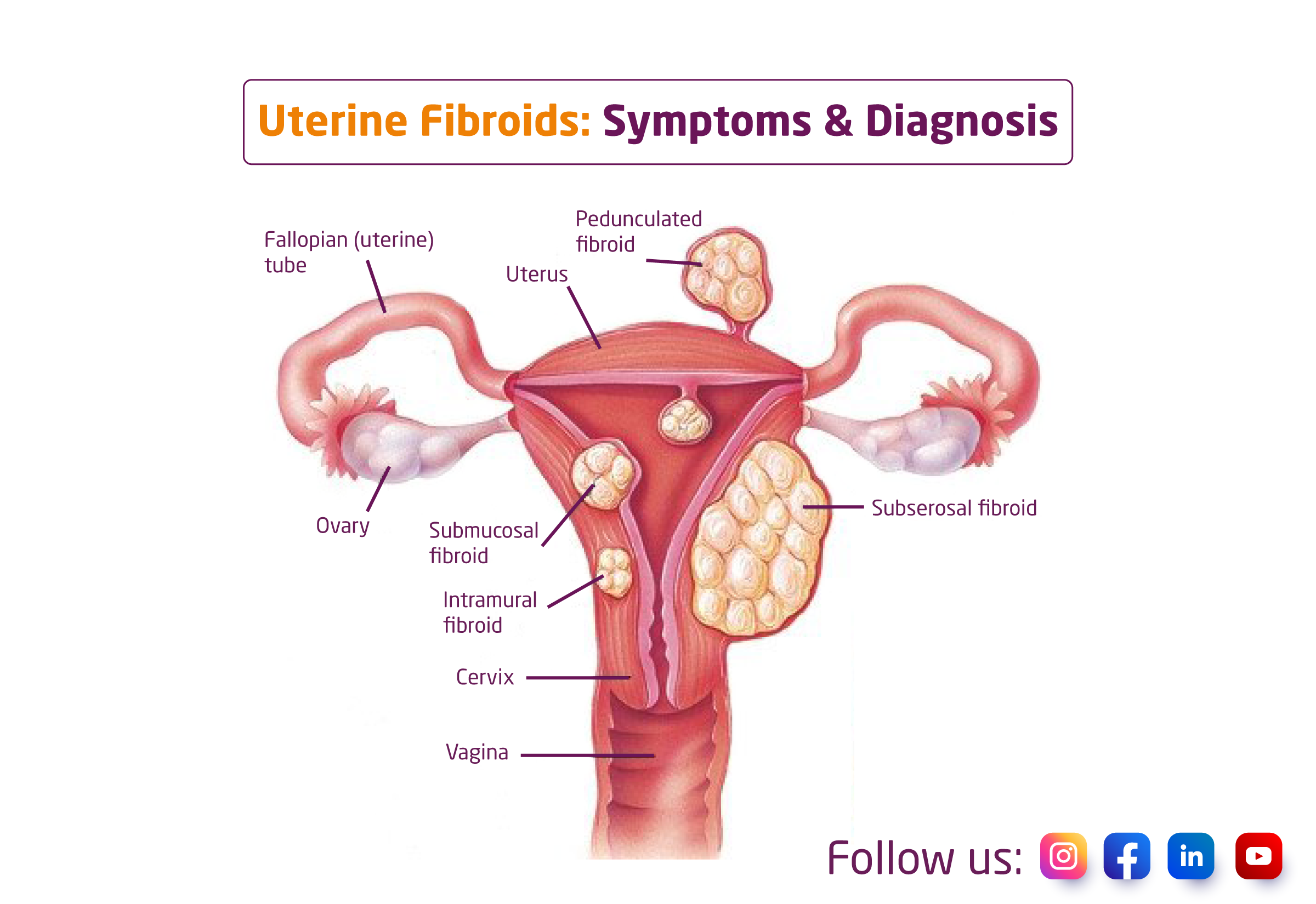
Leiomyomas, or uterine fibroids, are growths of muscle and connective tissue from the uterus’s wall. Often, these growths are not malignant (benign). In your pelvis, your uterus is an organ with a pear-shaped shape on its side. During pregnancy, the baby develops and grows inside the uterus.
Who is at risk for uterine fibroids?
Your likelihood of having fibroids may be influenced by a number of risk factors. They may consist of the following:
- Increase in body weight and obesity
- Family history of fibroids.
- Menopause at a late age.
- Getting your period at a young age.
- Not having children.
What are the symptoms of uterine fibroids?
Most fibroids are symptomless and don’t need any treatment beyond routine monitoring by your healthcare provider. Usually, these are little fibroids. Asymptomatic fibroids are those that do not cause symptoms. You may have a number of symptoms from larger fibroids, including:
- Excessive or uncomfortable menstrual bleeding.
- Bleeding in between menstrual cycles.
- Bloating or a lower abdominal fullness sensation.
- Frequent urination.
- Pain during sex.
- Low back pain.
- Constipation.
- Chronic vaginal discharge.
- Inability to totally empty your bladder or urinate.
- Increased abdominal enlargement.
How are uterine fibroids diagnosed?
Frequently, fibroids are initially identified during a routine visit with your doctor. These can be detected during prenatal care or a gynecologic exam and felt during a pelvic exam. Quite frequently, your description of severe bleeding and other relevant symptoms may prompt your doctor to think about including fibroids in the diagnosis. Several tests can be performed to identify the size and location of fibroids as well as to confirm their presence. These tests might be:
- Ultrasonography: This non-invasive imaging examination uses sound waves to paint a picture of your inside organs. Transvaginal or transabdominal ultrasonography procedures can be used.
- Magnetic resonance imaging (MRI): This examination produces finely detailed images of your interior organs using magnets and radio waves.
- Computed tomography (CT): Using X-ray imaging, a CT scan creates a detailed view of your inside organs from various angles.
- Hysteroscopy: A scope is a small, flexible tube with a camera on the end that your doctor will use during a hysteroscopy to view the fibroids inside your uterus. The scope is inserted into your uterus after passing through your cervix and vagina.
- Hysterosalpingography (HSG): In order to get a clear image of the uterus, a contrast substance must first be injected. This is more frequently applied to those who are also having infertility testing.
- Sonohysterography: In this imaging procedure, a tiny catheter is inserted transvaginally, and saline is administered into the uterine cavity via the catheter. With the help of the additional fluid, your uterus can be seen more clearly than during a typical ultrasound.
- Laparoscopy: Your doctor will create a tiny cut (incision) in your lower abdomen to do this test. To take a close-up look at your inside organs, a little, flexible tube with a camera on the end will be inserted.
